Views: 0 Author: Site Editor Publish Time: 2026-09-03 Origin: Site
Upgrading your imaging systems in 2026 unlocks a step-change in diagnostic confidence. You will visualize pathology earlier and with remarkable clarity, reducing ambiguous findings and repeat scans. New technology also cuts MRI scan times by up to 50%, boosting daily patient throughput despite flat reimbursement rates. This strategic investment future-proofs your practice against evolving clinical standards. Partner with a trusted imaging equipment supplier for ultrasound examination for sale and MRI Magnetic Resonance options that elevate both outcomes and revenue.
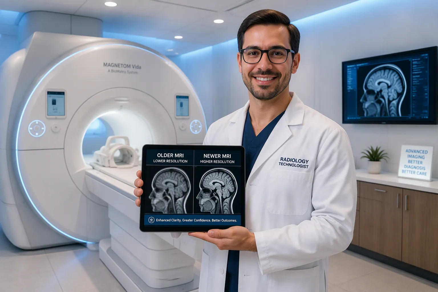
Upgrading to 2026 imaging systems improves image clarity, so doctors spot diseases earlier and more accurately.
New AI tools automate measurements and comparisons, saving 5 to 14 minutes per scan and boosting daily patient capacity.
Modern MRI machines are quieter and faster, cutting scan times by half and reducing patient anxiety for better results.
You see pathology earlier and with far greater clarity when you upgrade to 2026 imaging systems. Modern ultrasound and MRI equipment delivers image resolution that reveals subtle tissue changes, faint fractures, and small lesions that older systems routinely miss. This leap in diagnostic accuracy directly transforms how you plan treatments and manage patients. AI algorithms trained on vast datasets now identify patterns you might overlook during manual interpretation—faint pleural lines, hairline rib fractures, small cortical breaks. These algorithms improve lesion conspicuity, ensuring critical findings never escape your attention.
AI algorithms trained on large datasets consistently identify subtle patterns such as faint pleural lines, hairline rib fractures, and small cortical breaks. This improves lesion conspicuity, ensuring that findings are not overlooked during manual interpretation.
In emergency and trauma settings, radiologist fatigue and high workloads can lead to missed subtle fractures or pleural effusions, delaying treatment. AI tools flag these subtle pathologies, reducing the rate of overlooked findings compared to manual reading alone. Computer-aided detection and triage systems automatically escalate suspected fractures, pneumothorax, or pleural effusion to the top of your worklist, ensuring critical cases never get overshadowed by routine examinations.
Advanced imaging capabilities also directly influence your treatment planning and patient management. AI-driven models enhance diagnostic accuracy, streamline workflows, and facilitate personalized treatment strategies. For example, early detection of liver, skin, and prostate cancers improves procedural accuracy in radiotherapy and cardiology. Functional MRI allows adaptive focal boosting and personalized dosage, tailoring treatment based on real-time response. These capabilities optimize treatment planning and delivery, ultimately improving patient outcomes.
AI automation transforms your radiology workflow by handling repetitive tasks that consume valuable time. Automated quantification tools measure and annotate findings without manual calipers, and they compare current studies to prior exams automatically. The documented time savings are substantial:
Workflow task | Manual method | AI-automated method | Documented time saving |
|---|---|---|---|
Measurement documentation | Manual caliper tools | AI auto-measurement with annotation | 2–6 minutes per study |
Prior comparison | Manual retrieval and review | AI auto-comparison with deltas | 3–8 minutes per study |
These savings apply equally to ultrasound and MRI systems when you integrate AI quantification tools. You recover 5 to 14 minutes per study, allowing you to scan more patients each day or dedicate more time to complex cases.
AI-assisted acquisition also reduces operator-dependent variability. This means you get consistent measurements regardless of who performs the scan:
AI-assisted acquisition reduces repeat scans and supports guided capture, directly minimizing differences in how operators acquire images.
Automated measurement and structured reporting requirements standardize measurements across global hospital and imaging networks, ensuring uniform output regardless of operator.
Acquisition guidance provides AI prompts and quality feedback, helping non-specialist users capture usable images with fewer repeat attempts, reducing operator skill gaps.
Workflow standardization fits AI software into enterprise governance, ensuring bedside scans reach PACS with clear review steps, enforcing consistent measurement protocols.
Longitudinal disease monitoring applications use automated tumor measurement, lesion-volume tracking, and RECIST/iRECIST assessment to improve consistency across repeated studies. This automation removes manual measurement variability between operators. Follow-up scans compare against standardized, AI-generated metrics rather than operator-dependent manual calipers, giving you reliable data for treatment decisions.
You can cut MRI scan times by approximately 50% with advanced acceleration techniques available in 2026 systems. A peer-reviewed study on combined compressed sensing and parallel imaging in 7T MRI systems reported an average scan time reduction of about 50% across tested sequences, with no significant degradation in image quality metrics like contrast-to-noise ratio or noise. This means you complete a 30-minute exam in 15 minutes, or a 60-minute protocol in 30 minutes.
Faster scans directly benefit your patients. Shorter exam duration reduces motion artifacts, especially for patients who struggle to remain still—children, elderly individuals, or those in pain. You also improve patient throughput, scanning more patients per day without extending your operating hours. This efficiency gain compensates for flat reimbursement rates and helps you maintain financial sustainability.
The technology works by acquiring less data and using sophisticated algorithms to reconstruct high-quality images. Parallel imaging uses multiple receiver coils to speed up acquisition, while compressed sensing exploits image sparsity to reconstruct from fewer measurements. When combined, these techniques deliver the diagnostic information you need in half the time. You do not sacrifice image quality; you gain efficiency and patient comfort simultaneously.
Your choice of imaging equipment supplier determines the success of your entire upgrade initiative. A trusted partner brings more than hardware to your facility. They bring clinical expertise, financial flexibility, and ongoing technical support. When you evaluate an imaging equipment supplier for ultrasound examination for sale and MRI Magnetic Resonance systems, you must look beyond the initial price tag. The right partnership transforms your capital investment into a long-term operational advantage.
Patient anxiety during MRI scans presents a significant clinical challenge. Statistics show that 10-14.5% of standard MRI scans terminate prematurely due to claustrophobia, panic, or anxiety. These interruptions waste valuable scanner time and delay diagnosis. Modern MRI systems address this problem directly through quieter operation and wider bore designs.
Evidence Type | Evidence Content | How it Supports Query |
|---|---|---|
Statistical evidence | 10-14.5% of standard MRI scans are prematurely terminated due to claustrophobia, panic, or anxiety. | This high baseline rate of interruptions indicates that reducing noise can lower terminations. |
Direct statement | "Combining silent MRI with simple preparation steps can help you complete the scan without sedation in most cases." | Directly shows that quieter MRI reduces the need for sedation. |
Direct statement | "Patients who previously abandoned a standard MRI due to claustrophobia often complete a silent MRI scan without sedation on their first attempt." | Demonstrates that silent MRI enables completion without sedation, thus reducing interruptions. |
Clinical observation | Children benefit from the quieter environment, reducing fear and sensory overload, often avoiding the need for general anaesthesia. | Shows impact on sedation need for pediatric patients. |
Quieter MRI technology reduces acoustic noise by up to 90% compared to conventional systems. This dramatic reduction changes the patient experience completely. Patients who previously abandoned a standard MRI due to claustrophobia often complete a silent MRI scan without sedation on their first attempt. Children benefit especially from the quieter environment. The reduced fear and sensory overload often eliminate the need for general anesthesia in pediatric cases.
Wider-bore systems provide another layer of comfort. A 70-centimeter bore opening accommodates larger patients and reduces the confined feeling that triggers anxiety. You position patients more easily, and they remain calmer throughout the examination. This enhanced comfort directly translates to higher image quality. Research from multiple studies confirms that improved patient satisfaction reduces involuntary motion during scans. Less motion means fewer artifacts and sharper diagnostic images. You capture the detail you need on the first attempt, avoiding repeat examinations.
Modern imaging systems incorporate safety innovations that protect both patients and staff. Advanced dose management technologies reduce radiation exposure without compromising image quality. You maintain diagnostic confidence while fulfilling your duty to minimize patient risk. These systems use intelligent exposure controls that automatically adjust parameters based on patient size and anatomy.
Contrast agent safety has also improved substantially. Newer MRI systems require lower doses of gadolinium-based agents while delivering equivalent diagnostic information. You reduce the risk of nephrogenic systemic fibrosis and gadolinium retention concerns. Ultrasound contrast agents now offer improved safety profiles with fewer adverse reactions. These advancements matter particularly for patients who require repeated imaging studies over time.
Your imaging equipment supplier should provide comprehensive safety training as part of the upgrade package. They should document dose reduction protocols and help you implement them across your facility. The best suppliers offer ongoing education about emerging safety guidelines and regulatory requirements. This partnership ensures your team operates at the highest safety standards from day one.
A thorough return on investment analysis requires examining more than the purchase price. You must evaluate the total cost of ownership across the equipment's lifespan. Your imaging equipment supplier for ultrasound examination for sale and MRI Magnetic Resonance systems should help you model these costs accurately. The following table outlines critical factors you must consider when selecting your 2026 supplier.
Critical Selection Factor | Key Consideration for 2026 Supplier Choice |
|---|---|
Total Cost of Ownership (TCO) | Evaluate acquisition, operation, maintenance, and end-of-life costs; compare refurbished vs. new scenarios to avoid misleading price comparisons. |
Service & Maintenance Agreements | Require SLAs with defined response times, uptime guarantees, and MRI-specific coverage (cryogen management, magnetic field homogeneity). |
Regulatory & Cybersecurity Compliance | Verify supplier's ability to meet updated FDA rules, Joint Commission standards, and provide security patches for aging equipment. |
Technology Lifecycle Alignment | Ensure supplier's equipment matches modality-specific replacement cycles (MRI: 7–10 years; Ultrasound: 5–8 years) and supports end-of-life notifications. |
AI & Software Integration | Confirm supplier offers embedded AI tools (reconstruction, reporting) as recurring license fees, not hidden capital costs. |
Service agreements deserve particular attention during your negotiations. Traditional full service agreements often make response time management difficult. Partnership models offer more creative approaches and greater cost reduction potential. One healthcare technology director notes that negotiating glassware options within partnership agreements reduced costs and downtime by more than 50 percent. You should seek similar flexibility in your own agreements.
"Having a solution in place for glassware will also improve uptime for the systems since you won't be searching for tubes and will likely have some on-site or close by your hospital," Riehm points out. "We have managed to reduce costs and downtime by more than 50 percent since negotiating the glassware options of our partnership agreements."
View your supplier as a partner rather than a vendor. Industry experts emphasize this distinction repeatedly. Busdicker recommends working collaboratively with suppliers to achieve the shared goal of enhancing patient care without driving up costs. Bowers adds that investing in supplier-provided training improves on-site response times and delivers year-over-year cost savings. These partnerships create value that extends far beyond the equipment itself.
Full service agreements make response time management difficult; partnerships allow creative approaches and greater cost reduction.
View suppliers as partners, not vendors, and work collaboratively to enhance patient care without increasing costs.
Invest in supplier-provided training to improve on-site response times and achieve year-over-year savings.
When you navigate the upgrade process, start with a comprehensive needs assessment. Your supplier should evaluate your current equipment, patient volume, clinical specialties, and facility constraints. They should present options that align with your specific operational goals. A reputable imaging equipment supplier offering ultrasound examination for sale and MRI Magnetic Resonance systems will provide transparent pricing models. They will also offer flexible financing arrangements that match your budget cycle.
The upgrade timeline requires careful planning. Your supplier should coordinate installation, staff training, and workflow integration with minimal disruption to your clinical operations. They should provide temporary equipment coverage during the transition period. This planning prevents revenue loss during the upgrade window. Your partnership should include regular performance reviews after installation to ensure the equipment delivers the expected return on investment.
Upgrading your imaging equipment transforms referral patterns, patient satisfaction, and your financial health. View this upgrade as a critical investment in clinical excellence. Competitive differentiation now comes from AI integration, portable systems, and hybrid platforms. Philips partnered with icometrix for AI-driven MRI brain scans. ZEISS renewed research with MPFI for advanced brain imaging. Consult an imaging equipment supplier for ultrasound examination for sale and MRI Magnetic Resonance systems. This imaging equipment supplier for ultrasound examination for sale and MRI Magnetic Resonance systems will conduct a needs assessment aligned with your goals.
You present concrete ROI data: reduced scan times increase daily throughput, AI automation cuts radiologist reading time, and fewer repeat scans lower operational costs. These metrics translate directly into financial sustainability.
Your supplier coordinates phased installation and staff training during off-peak hours. They provide temporary equipment coverage during transition. Most facilities maintain full imaging capacity throughout the upgrade process with proper planning.
MRI systems typically serve 7–10 years; ultrasound equipment lasts 5–8 years. Your supplier should offer modular upgrades and software updates that extend system relevance. Choose equipment with AI-ready architecture to accommodate future innovations.